Vein Stripping

Sunday, June 28, 2009

Basic Facts

· Vein stripping means the excision, or removal, of large veins and the closing off of smaller vein tributaries.
· The removal of smaller veins with a technique called small incision avulsion is often done with vein stripping.
· Vein stripping is commonly used to improve the appearance of the legs and to relieve symptoms of varicose veins, unnaturally and permanently distended veins that lie directly under the skin.

The body has veins located close to the surface of the skin that help return oxygen-depleted blood to the heart. As superficial vein walls stretch and weaken because of age or heredity, blood can begin to flow backward and collect inside the veins. This in turn may cause increased pressure that can permanently damage the elastic vein walls.

When the walls of veins in the leg stretch and bloat out of shape, the valves that normally prevent blood from flowing backward also become distorted and the veins can begin to malfunction. Varicose veins or spider veins may result.

Varicose veins appear as blue, bulging and twisted veins, visible through the skin on a person’s legs. Left untreated, varicose veins can cause tiredness or heaviness in the legs. Affected areas of the leg may also ache or burn. In severe cases, varicose veins can lead to swollen ankles and scaly, dry skin.

The goals of vein stripping are to relieve pain, to improve circulation through the venous system by removing pathways of blood reflux and to improve the appearance of a person’s leg. Vein stripping involves the removal of the saphenous vein in the leg and any varicose tributary veins.

WHEN IS IT INDICATED?

Physicians decide to perform vein stripping after a complete duplex ultrasound evaluation of the veins shows that the veins are diseased and malfunctioning.

PRE-TREATMENT GUIDELINES

Vein stripping is typically an outpatient procedure, but it is commonly performed in the operating room using general or occasionally, spinal anesthesia.

WHAT TO EXPECT

After shaving a small site on the upper leg to help minimize the risk of infection, the physician makes a small incision near the groin to access the upper end of the great saphenous vein.

Once the physician can see the vein, he or she disconnects and ties off the main vein tributaries associated with it and disconnects the saphenous vein from the femoral vein. A stiff but flexible wire is inserted into the free end of the saphenous vein and advanced down its length and out through a second incision made at the upper calf, just below the knee.

The end of the vein nearest the groin is tied tightly, and then the knot is tied to the end of the wire, or a metal head is attached to the end of wire at the groin. Smaller veins connected to the saphenous vein along the leg may be cut away with tiny incisions, and then the physician pulls the wire downward. As the wire travels through the length of the vein it pulls the vein with it, turning the vein inside out and pulling it away from the smaller tributary veins.

The vein is removed through the incision in the upper calf, although some doctors may strip the vein all the way to the ankle. The incisions are closed with stitches and compression bandages are applied along the length of the leg.

POSSIBLE COMPLICATIONS

The risks of vein stripping are typically small, because the procedure is usually performed in people who are in generally good health. As in any vein surgery, blood clots are a special concern.

Certain complications can occur with any surgery requiring general anesthesia. They include:
· Allergic reactions;
· Problems breathing;
· Bleeding; and
· Infection.

Other complications are specific to vein stripping, including bruises, recurrence of varicose veins, and nerve injury.

POST-TREATMENT GUIDELINES

To control swelling and bleeding and promote healing of the surgical wounds, the treated areas may be firmly wrapped with elastic bandages. Compression stockings may also be used. Typically, a person’s legs will be firmly bandaged for 36 hours after vein stripping, and the legs are usually rewrapped and compression continued for days and even weeks after the procedure.

People who have had vein removal surgery may be prescribed a mild analgesic. The most important precaution in the postoperative period is taking 10 to 12 short walks per day, each lasting 5 to 10 minutes. Most patients are able to return to their usual daily activities within two weeks.

CONTACT INFORMATION

To schedule a visit with a Baylor physician, prospective patients should contact the Baylor Clinic at (713) 798-5700.

Maze Heart Surgery

Overview

Mayo Clinic is one of a few medical centers in the United States that performs the maze procedure, a highly complex surgical treatment option for atrial fibrillation. Mayo Clinic heart surgeons are highly skilled and have years of experience with this technique.

The maze procedure can cure atrial fibrillation by creating barriers to the electrical pathways, in the form of scar tissue, in the atria (the heart's upper chambers).

In the procedure, the surgeon creates multiple cuts into the atria muscle in an intricate pattern, or maze, and then stitches the incisions together to produce scars. Because the scars do not carry electrical signals, they interfere with stray electrical impulses that cause atrial fibrillation and, as a result, allow the heart to restore a regular, coordinated heartbeat.

Of Mayo Clinic patients who undergo the maze procedure for atrial fibrillation, 95 percent have no atrial fibrillation at discharge. But those who do not respond to the procedure may have more success with drug therapy afterward. Patients also benefit from Mayo's state-of-the-art diagnostic and treatment capabilities for patients with heart arrhythmias.

New technology is making the maze procedure faster, safer and less invasive. Mayo Clinic is involved in the research and use of new methods for the maze procedure, including:

Cryotherapy
At Mayo Clinic, surgeons routinely use cryotherapy to reduce the extent of surgical incisions made with a scalpel. In cryotherapy, surgeons use a device to freeze heart tissue and create lesions on the atria. Electrical barriers can be created in 60 to 90 seconds, minimizing the time to perform the maze procedure.

Minimal Access Catheter Maze Procedure
Physicians use catheter radiofrequency ablation (atrial fibrillation ablation) to replicate the lines of scar tissue that are created with the open-heart maze procedure surgery.

Mayo Clinic is an international leader in catheter ablation and uses the most advanced technology to treat arrhythmias. Read more about atrial fibrillation ablation.

Advantages of the Maze Procedure

  • Corrects atrial fibrillation; many patients require no further treatment
  • Restores a regular, coordinated heartbeat
  • For many patients, brings freedom from long-term use of blood-thinning medications
  • Lowers risk of developing blood clots or strokes
  • Decreases symptoms, such as fainting or near-fainting

Because the maze procedure requires open heart surgery, it is usually reserved for patients whose atrial fibrillation has not responded well to medication, whose symptoms interfere with their quality of life, and those at high risk for blood clots or stroke.

New Alternatives to Open Heart Surgery for Children

Open heart surgery is absolutely essential for the treatment of many types of congenital heart defects. However, there are an increasing number of defects that can be treated with special catheters or tiny devices that can be folded into catheters. If a defect can be treated effectively with a catheter technique, the procedure is much easier on the patient.

Most catheter treatment procedures are done with the patient sedated or under mild anesthesia. The only incision is a 1/8-inch insertion point over the blood vessels (usually in the legs). These do not even require a stitch. In most cases, patients are able to leave the hospital the same day as the procedure or sometimes, after an overnight observation period. Patients can return to school or to work within 1 to 2 days after a catheter treatment procedure in most cases. Because of the greatly shortened hospital stay and reduced need for complicated equipment in the operating room and intensive care unit, the costs of catheter procedures are usually significantly less than the cost of treatment of the same defect by surgical techniques.

Common catheterization procedures

The most common types of interventional catheterization procedures are those performed to:

  • Create septal defects
  • Open stenotic valves
  • Open stenotic vessels
  • Close abnormal vessels
  • Close certain septal defects
Creating septal defects

Certain types of congenital heart defects require that there be a large communication between the two atria (upper chambers of the heart). These heart conditions include transposition of the great vessels and some patients with mitral or aortic stenosis or atresia. The communication can be created by either rapidly pulling a round balloon through the defect to tear the septum or by placing a long valvuloplasty balloon in the septum and inflating it to a large size. These procedures are usually temporary procedures to allow the patient to recover or survive until the time of surgery.

Opening stenotic valves

Both the aortic and pulmonary valves can be treated in many patients with aortic or pulmonic stenosis. The basic technique is to advance a special catheter through the small opening in the valve. A long balloon on the catheter is then rapidly inflated and deflated. The procedure works by pulling apart the fused leaflets of the stenotic valve.

Pulmonary balloon valvuloplasty is almost universally considered the treatment of choice for isolated pulmonary stenosis. Balloon aortic valvuloplasty is a technically more difficult procedure to perform and carries a higher risk than pulmonary valvuloplasty so that only experienced centers utilize this technique.

The results with balloon valvuloplasty at The Cleveland Clinic Foundation have been excellent and this is usually the first procedure done on patients who do not have significant leakage of the aortic valve.

Opening stenotic vessels

Children with congenital heart disease frequently have narrowed vessels in the pulmonary arteries or may have coarctation of the aorta which is a narrowing or stenosis of the aorta.

Balloon angioplasty is the procedure wherein a special balloon catheter is advanced to the stenotic area. The balloon is rapidly inflated and deflated.

The procedure works by stretching the stenotic area enough that small tears are created in the inner two layers of the vessel wall. If the vessel is not dilated enough, then the stenotic area will simply stretch and return to it's original size. If the area is dilated too far, there is the possibility of rupture or creating a thin enough area in the vessel that an aneurysm forms later.

In older patients, stents can be used to overcome the elastic recoil of the vessel and usually provide a better result. Angioplasty procedures may need to be repeated and if stents are placed in growing children, they will need to be further dilated as the child grows larger.

Closing abnormal vessels

The most common abnormal vessel that need to be closed is the patent ductus arteriosus (PDA). If this vessel is large, then the patient may have signs of heart failure and will have a significantly shortened life.

If the PDA is small, the patient may have no symptoms, but may be at risk for an infection called endocarditis which is fatal if not treated with high doses of intravenous antibiotics for 4 to 6 weeks. Closing the PDA eliminates both of these problems. PDAs are most often closed with a device called a coil. This is a long, slinky-like device with long cloth fibers imbedded in it. Coils can be straightened and pushed through a small catheter. When they come out of the catheter, they form loops of a predetermined size. If these loops are very carefully placed in the PDA, then they will cause clumping of blood cells in the material and will form a plug to close the vessel. In over 90 percent of patients, PDAs can be closed with the catheter treatment. Surgery or a specially designed PDA closure device can be used to close larger PDAs.

Closing septal defects

Several devices have been devised to fold up into catheters and open like umbrellas to close atrial septal defects and certain types of other septal defects. These devices have proven to be effective in most ASD patients. We use the one device that has received FDA approval (Amplatzer ASD device) and also use some newer devices that may have some advantages but are still investigational. The ASD devices are usually placed with the aide of transesophageal echocardiography or intracardiac echocardiography. Most patients can leave the hospital later on the day of their procedure. Investigational devices to close a hole between the ventricles (VSD) may be used in some patients who are at high risk for surgery.

Minimally invasive heart surgery

Definition

Minimally invasive heart surgery refers to surgery performed on the beating heart to provide coronary artery bypass grafting. This technique is often referred to as MIDCAB, minimally invasive direct coronary artery bypass; or OPCAB, off-pump CABG.


Purpose

Minimally invasive heart surgery is performed on the diseased heart to reroute blood around clogged arteries and improve the blood and oxygen supply to the heart. This approach provides patients some benefit in that cardiopulmonary bypass (use of a heart-lung machine) may be avoided, and smaller incisions can be used instead of the standard sternotomy (incision through the sternum, or breast bone) approach. Faster recovery time, decreased procedure costs, and reduced morbidity and mortality are the goals of this technique.

Minimally invasive technique is not new to the field of cardiac surgery. It was performed as early as the 1950s, although the technology associated with stabilizing the cardiac structure during the procedure has become more sophisticated. Also, the anesthesiologist and perfusionist (person monitoring blood flow) have developed better techniques to preserve cardiac function during the procedure to help the surgeon achieve the desired outcome. During the 1990s these new techniques were named: off-pump CABG (OPCAB) and minimally invasive direct coronary artery bypass (MIDCAB). The MIDCAB procedure includes procedures done both with and without cardiopulmonary bypass, the later being referred to as off-pump MIDCAB. Unless otherwise specified, MIDCAB refers to both types of procedures.

Minimally invasive valve surgery has been an outgrowth of the success with minimally invasive coronary artery bypass grafting. Incisions other then the traditional sternotomy allow access to the heart. Minimally invasive valve surgery still requires cardiopulmonary bypass, since this is a true open-heart procedure, (i.e. this is not surgery that is done while the heart is beating). New tools in managing cardioplegic cardiac arrest allow for the smaller incision unobstructed by the required instrumentation. Cannulation of the femoral vessels instead of the larger vessels of the heart also improves visualization.


Demographics

Patients under the age of 70, but not limited by age, with a history of coronary artery disease can be evaluated for this procedure. High risk patients with advanced age, at risk for stroke, or suffering peripheral vascular disease, renal disease, or with poor lung function may benefit from OPCAB and MIDCAB.

Typically disease of the left anterior descending coronary artery is treated with the technique called off-pump

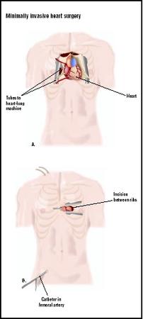
In traditional open heart surgery, a large incision is made in the chest, and the sternum must be broken (A). Minimally invasive surgery uses a much smaller incision between the ribs to access the heart (B). (Illustration by GGS Inc.)
In traditional open heart surgery, a large incision is made in the chest, and the sternum must be broken (A). Minimally invasive surgery uses a much smaller incision between the ribs to access the heart (B). (
Illustration by GGS Inc.
)

MIDCAB. With sternotomy, disease of the right and left coronary arteries can also be addressed by OPCAB. The significance and location of the coronary artery lesions may limit the success of the MIDCAB or OPCAB procedure. Most practices have at least one surgeon skilled in performing revascularizations without cardiopulmonary bypass. Of all coronary artery bypass grafting procedures, approximately 10–20% are performed in this manner.

Description

The patient receives cardiac monitoring during general anesthesia. Systemic anticoagulation is given to avoid clot formation from foreign surfaces and any periods of artery blockage (occlusion).


MIDCAB

If cardiopulmonary bypass is not employed, the procedure is called an off-pump MIDCAB. The surgeon performs an alternative incision (rather than a midline sternotomy), typically a left anterior thoracotomy. The left internal mammary artery is dissected from the left chest wall. A stabilizer device is placed on the heart to provide support of the left anterior descending artery as the heart continues to beat. This device applies gentle pressure or suction, mildly limiting cardiac function. The left internal mammary artery is sutured to the left anterior descending artery to bypass the blockage (anastomosis).

If cardiopulmonary bypass is indicated, the surgeon inserts cannulae (small, flexible tubes) into the femoral vessels. Aortic occlusion and cardioplegia are administered through a catheter advanced through the contralateral femoral artery into the aortic root (ascending aorta). This catheter has a balloon tip that stops blood flow to the coronary arteries when inflated, but allows selective administration of cardioplegia (a solution that stops the heart) to the coronary arteries. Angiography is performed to provide visualization of catheter placement.

The surgeon performs an alternative incision (rather than a midline sternotomy), typically a left anterior thoracotomy. The left internal mammary artery is dissected from the left chest wall. Cardiopulmonary bypass can be instituted with or without cardioplegic arrest. Cardioplegic arrest requires cardiopulmonary bypass. The use of cardioplegic arrest makes this a non-beating heart procedure, but it is still considered MIDCAB. Cardioplegic arrest of the heart occurs as the balloon tip of the catheter is inflated. The left internal mammary artery is sutured to the left anterior descending artery to bypass the blockage (anastomosis). Once the anastomosis is complete the balloon is deflated, allowing the heart to begin to beat. Cardiopulmonary bypass is discontinued once cardiac function is stabilized. The cannulae and catheter are removed, and the groin wounds are closed with sutures.

OPCAB

The OPCAB procedure does not use cardiopulmonary bypass. The incision of choice can be a midline sternotomy or a left anterior thoracotomy (incision into the side). The midline sternotomy allows access to both the right and left internal mammary arteries. Additional vascular bypass conduits may be acquired by harvesting the saphenous vein (in the leg), gastroepiploic artery (near the stomach), or radial artery (in the arm). A stabilizing device is used to secure the coronary artery of choice. This device applies gentle pressure or suction, mildly limiting cardiac function, but providing better access to posterior and inferior vessels of the heart. The surgeon makes the necessary anastomosis to the targeted coronary arteries. If conduits other then the mammary arteries are used they are connected to the ascending aorta to provide systemic blood flow.

If an anticoagulant was administered, drugs are given to reverse the anticoagulant. Upon completion of the off-pump MIDCAB, MIDCAB, or OPCAB procedure, the chest is closed. If a midline sternotomy was performed, stainless steel wires are implanted to hold the sternal bone together. Sutures are used to close the skin wound, and sterile bandages are applied as a wound dressing.


Diagnosis/Preparation

An electrocardiogram detects the presence of acute coronary blockage (occlusion). A history of myocardial infarction can also be detected by electrocardiogram. Patients with a history of angina also are evaluated for coronary artery disease. Coronary angiography provides the best diagnostic information about the extent and location of the coronary artery disease.

Aftercare

The patient receives continued cardiac monitoring in the intensive care unit. Once the patient is able to breathe on his/her own, the breathing tube is removed (extubation), if it is not removed immediately post-operatively. Any medications to treat poor cardiac function or manage blood pressure are discontinued as cardiac function improves and blood pressure stabilizes. Blood drainage tubes protruding from the chest cavity are removed once internal bleeding decreases. The patient also may be equipped with external cardiac pacing to maintain heart rate. The pacing is terminated once the heart is beating at an adequate rate free of arrhythmia. A warming blanket may be used to warm the patient's core temperature that was decreased by the surgical exposure.

The duration of the post-operative hospital stay is reduced by one to two days in these procedures. Pain also should be reduced. Homecare for the wound is described prior to discharge, and instructions for responding to adverse events after discharge also are given. Patients who have undergone these procedures should expect to return to normal activities sooner than those who have undergone traditional coronary artery bypass grafting.


Risks

MIDCAB can result in a higher rate of restenosis (recurrence of narrowing of the arteries) then traditional coronary artery bypass grafting, but these numbers continue to decrease as experience with the procedure improves. Some patients may have to have the procedure converted to a standard sternotomy with cardiopulmonary bypass, if the anastomosis can not be completed from the MIDCAB approach. Rib fracture is the most common adverse event. Pericarditis also is a possible complication. Supraventricular arrhythmias and ST segment elevation also may develop.

In the event of systemic blood pressure abnormalities, arrhythmia, poor surgical anastomosis, or poor exposure of the coronary blood vessels, OPCAB patients may require conversion to cardiopulmonary bypass for completion of the anastomosis. Post-operatively some patients may need additional surgery to control bleeding or to address poor sternal healing. This is related to the increased use of both internal mammary arteries for these procedures. Cerebral complications and atrial fibrillation also may be experienced. These post-operative complications are comparable to those seen in patients who have undergone traditional coronary artery bypass grafting.


Normal results

Patency (openess) of the grafted vessels is expected to be the same as what is seen in traditional coronary artery bypass grafting. When compared to traditional coronary artery bypass grafting, minimally invasive heart surgery also is expected to result in a shorter hospital stay, less pain, fewer blood transfusions, and quicker return to normal activity.

Morbidity and mortality rates

MIDCAB

Conversion to a full sternotomy or sternotomy with cardiopulmonary bypass is expected in 1–2% of patients. Redo procedures and reoperation can occur in over 5% of patients, which is still lower than the risk of a second procedure associated with balloon angioplasty and stent placement. Over 90% of all patients are expected to be free of adverse events. Complications most frequently involve rib fracture (over 10% of patients). Mortality associated with MICAB is low and is not seen during the surgical procedure in most instances, but is associated with post-operative complications.


OPCAB

Conversion to cardiopulmonary bypass may be required in patients if anastomosis cannot be completed due to unstable blood pressure, arrhythmia, ischemia, poor anastomosis, or poor surgical access. The same operative mortality is expected when compared to cardiopulmonary bypass patients. The expected decrease in neurological events, renal dysfunction, pulmonary complications, or arrhythmias has not yet been shown to be a consistent benefit, therefore all of these complications can still occur.


Alternatives

Percutaneous balloon angioplasty and coronary stenting of the left anterior descending artery are successful alternative procedures. MIDCAB may be a preferred treatment when compared to balloon angioplasty and stenting because fewer repeat interventions are required. An additional alternative is traditional on-pump, cardiopulmonary bypass; coronary artery bypass grafting is a powerful technique with a long record of safety and effectiveness since the 1960s.


Resources

BOOKS

Hensley, Frederick A., Donald E. Martin, and Glenn P. Gravlee, eds. A Practical Approach to Cardiac Anesthesia. 3rd ed. Philadelphia: Lippincott Williams & Wilkins 2003.

PERIODICALS

Borst, H. G. and F. W. Mohr. "The History of Coronary Artery Surgery—A Brief Review." The Thoracic and Cardiovascular Surgeon 49 (2001): 195–198.

Holubkov, R., et al. "MIDCAB Characteristics and Results: the CardioThoracic Systems (CTS) Registry." European Journal of Cardio-Thoracic Surgery 14, suppl.1 (1998): S25–S30.

Lund, O., et al. "On-pump Versus Off-pump Coronary Artery Bypass: Independent Risk Factors and Off-Pump Graft Patency." European Journal of Cardio-Thoracic Surgery 20 (2001): 901–907.

Moussa, I., et al. "Frequency of Early Occlusion and Stenosis in Bypass Grafts After Minimally Invasive Direct Coronary Arterial Bypass Surgery." The American Journal of Cardiology 88 (2001): 311–313.


Allison Joan Spiwak, MSBME

WHO PERFORMS THE PROCEDURE AND WHERE IS IT PERFORMED?


Medical centers performing cardiac surgical procedures are equipped to perform this procedure. A cardiothoracic, cardiovascular, or cardiac surgeon receives additional training to successfully complete this procedure. Special technology in stabilizer design is purchased by the institution and made available for the surgeon to master. Within most clinical practices one surgeon becomes skilled in the technique. This one surgeon completes most procedures off-pump with MIDCAB or OPCAB techniques as necessary to revascularize the patient.

QUESTIONS TO ASK THE DOCTOR


  • Is there a surgeon associated with this practice skilled with OPCAB or MIDCAB procedures?
  • Can the surgeon skilled in these procedures evaluate the patient for an OPCAB or MIDCAB procedure?
  • How many procedures has the surgeon performed in the last year? In the last five years?
  • What is the surgeon's reoperation rate in regards to length of graft patency?

Alternative To Open Heart Surgery Interventional Cardiologists Help The Faint Of Heart Without Surgery

January 1, 2009 — Interventional cardiologists created an alternative to open heart surgery by developing a mitral valve clip. To alleviate mitral valve regurgitation--a condition where the heart's mitral valve does not close properly, allowing blood to leak back into the heart--cardiologists insert a catheter into the patient's groin that travels up into the mitral valve. The clip is fed through this catheter, where it finally grasps and tightens the valves' leaflets--effectively preventing blood from leaking. The clip remains in place while the catheter is removed, the entire procedure taking approximately two hours and recovery a few weeks. The procedure is good for those with weaker hearts, when traditional surgery is more dangerous.

Chances are you know someone who has had heart problems. In fact, one in five people over the age of 55 has a problem with their mitral valve. A new alternative to open heart surgery can get their blood flowing again.

Nothing keeps 77-year-old Josephine Herndon from shopping, but her hobby was slowed down by a heart problem called mitral regurgitation.

"In the store, I sat down, and I was breathing pretty heavily," said Herndon. "I could barely make it back to the car."

Mitral regurgitation is a condition in which the heart's mitral valve doesn't close tightly, allowing blood to flow backward into the heart.

"I had a leaky valve and didn't even know it," Herndon said.

"A lot of these patients have shortness of breath," said George Hanzel, M.D., an interventional cardiologist at William Beaumont Hospital in Royal Oak, Mich. "The main thing they have is fatigue, exercise intolerance, shortness of breath and swelling."

Herndon was one of the first patients in the United States to have the mitral clip procedure. First, interventional cardiologists inserted a catheter into her groin up into the mitral valve. Next, a clip was fed through. The clip grasped and tightened the valves' leaflets, preventing blood from leaking.

"By pulling them together and approximating them, it reduces the leakiness," Dr. Hanzel said.

The clip stays and keeps blood from leaking, and the catheter is removed. The procedure takes two hours, the same as for open-heart surgery. The difference is in the recovery -- down from months to just weeks.

"Patients typically say they feel better," Dr. Hanzel said. "They can breathe better. They can do more without having to stop and rest."

Herndon's mitral regurgitation was reduced from severe to trivial, and she's back looking for bargains again. "I always did love to go shopping," Herdon said.

The mitral clip procedure is good for patients who have a weak heart and may not make it through traditional surgery. The procedure is being investigated in clinical trials in 38 hospitals across the country.

ABOUT MITRA CLIP: The Mitra Clip is a device inserted into the heart by a catheter. It is used to gather and fasten the leaflets of the mitral valve of the heart, which can become loose enough to allow blood to leak when the valve is closed. Doctors insert the catheter into the femoral artery, and then work it through the body to the heart. Using this technique can help patients recover more quickly from mitral valve repairs.

HAVE A HEART: The heart pumps 5.6 liters of blood through the entire body in roughly 20 seconds; each day your blood travels some 12,000 miles, and your heart beats about 100,000 times. This delivers oxygen and other essential nutrients to the body's cells and organs. A heart attack occurs when the blood supply to the heart muscle is cut off, either because part of the heart is damaged (such as the valves to the chambers), or because plaque has built up inside the arteries, narrowing them and severely restricting blood flow. Symptoms of a heart attack include a squeezing discomfort in the center of the chest, pain or tingling in the left arm, shortness of breath and sometimes a cold sweat, nausea, or dizziness.

ABOUT HEART DISEASE: Most heart diseases arise from hardening of the arteries, especially from the buildup of fatty material along the inner lining of the arteries. Coronary arteries supply blood to the heart. When a blockage occurs, this flow is decreased. Heart medications target these blockages in several different ways. Nitrates dilate the veins, decreasing the oxygen requirements of the heart. They also dilate the coronary arteries to increase blood flow to the heart. Beta-blockers decrease the heart rate and the force of the heart's contractions. Aspirin prevents platelets in the blood from clotting and clumping on blood vessel walls.

Women's Heart Attack Symptoms Different from Men's

Research by the National Institutes of Health (NIH) indicates that women often experience new or different physical symptoms as long as a month or more before experiencing heart attacks.

Among the 515 women studied, 95-percent said they knew their symptoms were new or different a month or more before experiencing their heart attack, or Acute Myocardial Infarction (AMI). The symptoms most commonly reported were unusual fatigue (70.6-percent), sleep disturbance (47.8-percent), and shortness of breath (42.1-percent)

.Many women never had chest pains
Surprisingly, fewer than 30% reported having chest pain or discomfort prior to their heart attacks, and 43% reported have no chest painduring any phase of the attack. Most doctors, however, continue to consider chest pain as the most important heart attack symptom in both women and men.

The NIH study, titled "Women's Early Warning Symptoms of AMI," is one of the first to investigate women's experience with heart attacks, and how this experience differs from men's. Recognition of symptoms that provide an early indication of heart attack, either imminently or in the near future, is critical to forestalling or preventing the disease.

In a NIH press release, Jean McSweeney, PhD, RN, Principal Investigator of the study at the University of Arkansas for Medical Sciences in Little Rock, said, "Symptoms such as indigestion, sleep disturbances, or weakness in the arms, which many of us experience on a daily basis, were recognized by many women in the study as warning signals for AMI. Because there was considerable variability in the frequency and severity of symptoms," she added, "we need to know at what point these symptoms help us predict a cardiac event."

Women's symptoms not as predictable
According to Patricia A.Grady, PhD, RN, Director of the NINR, "Increasingly, it is evident that women's symptoms are not as predictable as men's. This study offers hope that both women and clinicians will realize the wide range of symptoms that can indicate heart attack. It is important not to miss the earliest possible opportunity to prevent or ease AMI, which is the number one cause of death in both women and men."

The women's major symptoms prior to their heart attack included:[/br]

  • Unusual fatigue - 70%
  • Sleep disturbance - 48%
  • Shortness of breath - 42%
  • Indigestion - 39%
  • Anxiety - 35%

    Major symptoms during the heart attack include:

  • Shortness of breath - 58%
  • Weakness - 55%
  • Unusual fatigue - 43%
  • Cold sweat - 39%
  • Dizziness - 39%

    Related NIH research into heart attacks in women includes possible ethnic and racial differences.

  • Women and Heart Failure

    Heart failure is a major cause of death and disability in women. Most cases of heart failure develop as a result of coronary artery disease or longstanding hypertension, and in most cases the causes, symptoms, and treatment of heart failure are similar in women and in men. You can read here about the causes, evaluation and treatment of heart failure. It is particularly important for women with heart failure to know this information, because the majority of women are NOT receiving optimal therapy for heart failure from their doctors, and that inadequate therapy leads directly to increased mortality and disability.

    In addition, some aspects of heart failure are different in women than in men. These differences include:

    Diastolic heart failuretends to be much more common in women than in men, and accounts for at least half the cases of heart failure in women. This condition is often missed by unalert physicians, and consequently women with diastolic heart failure often do not receive optimal therapy.

    Women can develop an acute condition called stress cardiomyopathy, or "broken-heart syndrome." While this condition appears rare, it does seem to affect women far more often than men.

    It is now demonstrated fairly convincingly that women tend to minimize their symptoms of heart failure, so that doctors are unaware of just how sick and disabled they actually are. This minimization of symptoms is one of the factors that leads to inadequate treatment in women with heart failure.

    Finally, evidence exists that treatment with digoxin may be dangerous in women. Digoxin is still one of the more common medications used in treating heart failure, but in many women its potential benefits may be outweighed by the risks

    Ten Things Your Surgeon Needs to Know

    When planning a surgery it is important to find out as much information as you can from your surgeon, but it is also essential that you give your surgeon all of the information needed to make your surgery as safe as possible. Here are ten things you absolutely must discuss with your surgeon in order to have a safe and healthy outcome.

    1. Medications

    Your surgeon needs to know about all of the medications you are taking, including prescription, non-prescription drugs, herbal supplements and vitamins. Supplements are often overlooked when listing current medications, but it is very important that the surgeon is aware of any supplements as they can interact with anesthesia and may increase bleeding.

    2. Smoking Habits

    Patients should be sure to notify their surgeon if they smoke, or have smoked in the past. Some smokers require more time to be taken off of the ventilator and supplemental oxygen once they are breathing on their own. Smoking can also impair wound healing and cause greater scarring than non smokers experience.

    3. Alcohol Intake

    It is essential that patients are candid about the amount of alcohol they consume. Patients who are dependent upon alcohol can have issues ranging from tremors to seizures as they begin to experience withdrawal. If the surgeon is aware that the patient is chemically dependent upon alcohol they can prescribe medications that will relieve the symptoms and prevent some of the more serious complications.

    Patients who are dependent upon alcohol may also have difficulty with pain control, as they are typically less sensitive to pain medication and require larger doses. If the surgeon is unaware of the alcohol use, the prescribed dosage may be inadequate.

    4. Previous Illnesses and Surgeries

    Surgeries leave scars, both internal and external, and can change surgeries that follow. A surgeon should be well aware of any previous surgeries, especially those that take place in the same region of the body. In addition to surgeries, any major illnesses should be disclosed as well, as a patient’s tolerance of anesthesia can be changed by previous and current illnesses.

    5. Illicit Drug Use

    Drugs, both prescription and illicit, can alter the way anesthesia effects patients. In addition, smoking illicit drugs, like smoking cigarettes, can alter the way a patient returns to breathing on their own after being on a ventilator.

    Illegal drugs can change the effectiveness of prescription pain medications, requiring different dosages and can have interactions with anesthesia drugs, causing serious complications.

    6. Allergies

    It is important to disclose all known allergies prior to having surgery. All allergies, including food, medications and those that cause skin irritation, should be included. By placing this information on your hospital chart, it will make the various departments of the hospital, including pharmacy and nutritional services, aware of the allergies.

    A good example is an egg allergy, which may not seem important when having surgery; however, many medications are formulated in an egg base, which could cause a serious reaction if given to the patient.

    7. Past Issues With Surgery

    The surgeon should be made aware with any problems with previous surgeries. This includes bleeding issues after surgery, briefly waking during surgery or anything else that was unusual. A patient who has had problems in the past is not necessarily going to have the same problems if they have surgery again, and problems that can reoccur may be prevented if the surgeon and anesthesia provider are aware of the issues.

    8. Current Illness or Fever

    If a patient begins to feel ill, or has a fever, in the days preceding surgery, the surgeon needs to be made aware. The surgeon may decide it is safe to continue with surgery or may opt to postpone the procedure. A fever is a sign of possible infection and should be disclosed, to prevent wasted time and energy for both the patient and the surgeon.

    A patient who presents at the hospital for a scheduled surgery unaware that they have a fever may be sent home and the surgery appointment changed.

    9. Current Health Conditions

    Any current health issues a patient is facing should be disclosed to the surgeon. For example, a patient who is having a knee replacement surgery needs to make their surgeon aware of the fact that they are diabetic and using insulin. Without this information, the hospital is unable to provide care for all of the conditions, which could harm the patient.

    10. Religious Issues

    Some religions forbid blood transfusions and other medical procedures. If this is the case, the surgeon must be aware of the conditions under which they are operating prior to surgery. Some surgeries would not be able to take place if the religious objection would impact the level of care. In other cases, there may be alternatives that the surgeon would be able to prepare if given enough time.

    What Happens During Open Heart Surgery

    n Pump Open Heart Bypass Surgery:

    An open heart bypass surgery is performed under general anesthesia, which requires that the patient be on a ventilator during surgery.

    Surgery begins with harvesting the blood vessels that will become the grafts. The saphenous vein in the leg is commonly used because it is long enough to create multiple grafts. If the saphenous vein cannot be used, vessels from the arm can be used instead. The left internal mammary artery is used for a single graft and is taken once the chest is opened for surgery.

    Once the saphenous vein has been recovered, the chest is opened by making an incision along the sternum, or breastbone. The surgeon then cuts the sternum, allowing the chest cavity to be opened, giving the surgeon access to the heart.

    In the traditional CABG procedure, the heart is stopped with a potassium solution so the surgeon is not attempting to work on a moving vessel, and the blood is circulated by a heart-lung machine. At this time the heart-lung machine does the work of the heart and the lungs, and the ventilator is not used.

    The surgeon places the grafts, either rerouting blood around the blockage, or removing and replacing the blocked vessel. The amount of time on the heart-lung bypass machine is determined by the speed at which the surgeon is able to work, primarily, how many grafts are needed.

    Once the grafts are complete, the heart is started and provides blood and oxygen to the body. The sternum is returned to its original position and closed using surgical wire, to provide strength the bone needs to heal, and the incision is closed.

    Pros of On Pump Open Heart Surgery:

    • Surgeon can operate quicker because the heart is still
    • Very little blood makes surgery faster
    • Appropriate for unstable patients

    Cons of On Pump Open Heart Surgery:

    • Increased inflammation/clotting after surgery
    • Transfusion more likely after surgery than with off pump
    • More fluid retention than off pump
    • Higher risk of kidney damage than off pump
    • Longer hospital stay than off pump
    • Increased risk of stroke

    Off Pump Open Heart Surgery:

    The procedure for beating heart, or “off-pump” surgery is essentially the same as the on pump surgery, but the flow of blood through the body is maintained by the heart during the procedure. Instead of using a heart-lung machine, the heart continues to beat but the area being grafted is held still by surgical instruments. Approximately 20% of first time CABG patients have off pump surgery.


    Pros of Off Pump Open Heart Surgery:

    • The heart is moving, slowing surgery
    • Less blood loss and fewer transfusions
    • Decreased risk of stroke
    • Decreased length of hospital stay
    • Less expensive

    Cons of Off Pump Open Heart Surgery:

    • Up to 70% of patients ineligible due to anatomy or medical condition
    • Rarely performed on an unstable patient

    Heart Surgery Overview

    Preparing for your heart surgery (video)
    Preparing for Your Heart Surgery
    (15 minute video)

    Thousands of heart surgeries are performed every day in the United States. In fact, in 2005 alone, surgeons performed 575,000 coronary bypass or valve repair and replacement surgeries. And even though there is a shortage of donor organs, in 2006, almost 2,200 people had heart transplants.

    Years ago, many doctors thought that heart surgery was a dream. Surgeons during World War II had learned how to operate on the heart, but they could not carry out what they had learned because it was hard to operate on a beating, moving heart. Also, the heart could not be stopped for more than a few minutes without causing brain damage.

    Two major advances in medicine made heart surgery possible:

    • The heart-lung machine, which takes over the work of the heart.
    • Body cooling techniques, which allow more time for surgery without causing brain damage.

    What is a heart-lung machine?

    perfusion_web

    Perfusion technologists operate the heart-lung machine during an open heart surgery procedure.

    The heart-lung machine is also called a cardiopulmonary bypass machine. It takes over for the heart by replacing the heart's pumping action and by adding oxygen to the blood. This means that the heart will be still for the operation, which is necessary when the heart has to be opened (open heart surgery). Because the heart-lung machine takes over the work of the heart, surgeons can operate on a heart that is not moving or full of blood.

    When you are connected to the heart-lung machine, it does the same job that your heart and lungs would do. The heart-lung machine carries blood from the upper-right chamber of the heart (the right atrium) to a special reservoir called an oxygenator. Inside the oxygenator, oxygen bubbles up through the blood and enters the red blood cells. This causes the blood to turn from dark (oxygen-poor) to bright red (oxygen-rich). Then, a filter removes the air bubbles from the oxygen-rich blood, and the blood travels through a plastic tube to the body's main blood conduit (the aorta). From the aorta, the blood moves throughout the rest of the body.

    The heart-lung machine can take over the work of the heart and lungs for hours. Trained technicians called perfusion technologists (blood flow specialists) make sure that the heart-lung machine does its job properly during the surgery. Even so, surgeons still try to limit the time that patients must spend hooked up to the machine.

    What are cooling techniques?

    Cooling techniques let surgeons stop the heart for long periods without damaging the heart tissue. Cool temperatures avoid damage to the heart tissue by reducing the heart's need for oxygen.

    The heart may be cooled in 2 ways:

    • Blood is cooled as it passes through the heart-lung machine. In turn, this cooled blood lowers body temperature when it reaches all of the body parts.
    • Cold salt-water (saline) is poured over the heart.

    After cooling, the heart slows and stops. Injecting a special potassium solution into the heart can speed up this process and stop the heart completely. The heart is then usually safe from tissue injury for 2 to 4 hours.

    View of a coronary artery bypass operation from observation dome overhead.

    View of a coronary artery bypass operation from observation dome overhead.

    Who is in the operating room during surgery?

    During heart surgery, a highly trained group works as a team. Here is a list of people who will be in the operating room during surgery.

    • The cardiovascular surgeon, who heads up the surgery team and performs the key parts of the surgery.
    • The assisting surgeons, who follow the direction of the cardiovascular surgeon.
    • The cardiovascular anesthesiologist, who gives you the medicines that make you sleep during the surgery (called anesthesia). The anesthesiologist makes sure that you get the right amount of medicines throughout the surgery and monitors the ventilator, which breathes for you during surgery.
    • The perfusion technologist, who runs the heart-lung machine.
    • The cardiovascular nurses, who are specially trained to assist in heart surgery.

    What kinds of heart and blood vessel surgeries are there?

    Many kinds of surgery are now performed on the heart and blood vessels.

    Coronary Artery Bypass

    This is the most common kind of heart surgery. You may also hear it called coronary artery bypass graft surgery (CABG), coronary artery bypass (CAB), coronary bypass, or bypass surgery.

    The surgery involves sewing a section of vein from the leg or arteries from the chest or another part of the body to bypass a part of a diseased coronary artery. This creates a new route for blood to flow, so that the heart muscle will get the oxygen-rich blood it needs to work properly.

    During bypass surgery, the breastbone (sternum) is divided, the heart is stopped, and blood is sent through a heart-lung machine. Unlike other kinds of heart surgery, the chambers of the heart are not opened during bypass surgery.

    When you hear the words single bypass, double bypass, triple bypass, or quadruple bypass, it refers to the number of arteries that are bypassed. The number of bypasses does not necessarily indicate how severe the heart condition is.

    See also on this site: Coronary Artery Bypass Surgery

    Valve repair or replacement

    Blood is pumped through your heart in only one direction. Heart valves play key roles in this one-way blood flow, opening and closing with each heartbeat. Pressure changes behind and in front of the valves allow them to open their flap-like "doors" (called cusps or leaflets) at just the right time, then close them tightly to prevent a backflow of blood.

    Two of the most common kinds of valve problems that require surgery are

    • Stenosis, which means the leaflets do not open wide enough and only a small amount of blood can flow through the valve. Stenosis occurs when the leaflets thicken, stiffen, or fuse together. Surgery is needed to either open the valve that is there or replace it with a new one.
    • Regurgitation, which is also called insufficiency or incompetence, means that the valve does not close properly and blood leaks backward instead of moving in the proper forward direction. Surgery is needed to either tighten or replace the valve.

    Surgical repair of a valve involves the surgeon rebuilding the valve so that it will work properly. Valve replacement means that the valve is replaced with a biological valve (made of animal or human tissue) or a mechanical valve (made from materials such as plastic, carbon, or metal).

    See also on this site:

    Arrhythmia Surgery

    Any irregularity in your heart's natural rhythm is called an arrhythmia. Arrhythmias are usually treated first with medicines. Other treatments may include

    • Electrical cardioversion, where the cardiologist or surgeon uses paddles to "shock" the heart back into a normal rhythm.
    • Catheter ablation, where the cardiologist uses a special tool to destroy (ablate) the cells that are causing the arrhythmia. This is done in the cardiac catheterization laboratory (the cath lab).
    • Pacing and rhythm-control devices, including pacemakers and implantable cardioverter defibrillators (ICDs).Patients can have these devices implanted while in the operating room or the cath lab.

    When these treatments do not work, surgery may be needed. One type of surgery is called Maze surgery. In Maze surgery, surgeons create a "maze" of new electrical pathways to let electrical impulses travel easily through the heart. Maze surgery is used most often to treat a type of arrhythmia called atrial fibrillation. Atrial fibrillation is the most common type of arrhythmia.

    See also on this site:

    Aneurysm Repair

    An aneurysm is a balloon-like bulge in a blood vessel or in the wall of the heart. An aneurysm occurs when the wall of a blood vessel or the heart becomes weakened. Pressure from the blood forces it to bulge outward, forming what you might think of as a blister. An aneurysm can often be repaired before it bursts.

    Surgery involves replacing the weakened section of blood vessel or heart with a patch or artificial tube (called a graft).

    Aneurysms in the wall of the heart occur most often in the lower-left chamber (called the left ventricle). These aneurysms are called left ventricular aneurysms, and they may develop after a heart attack. (A heart attack can weaken the wall of the left ventricle.) If a left ventricular aneurysm leads to an irregular heartbeat or to heart failure, the surgeon may perform open heart surgery to remove the damaged part of the wall.

    See also on this site:

    Transmyocardial Laser Revascularization (TMLR)

    Angina is the pain you feel when a diseased vessel in your heart (called a coronary artery) can no longer deliver enough blood to a part of the heart to meet its need for oxygen. The heart's lack of oxygen-rich blood is called ischemia. Angina usually occurs when your heart has an extra need for oxygen-rich blood, such as during exercise. Angina is nearly always caused by coronary artery disease (CAD).

    Transmyocardial laser revascularization (TMLR) is a procedure that uses lasers to make channels in the heart muscle, in an attempt to allow blood to flow from a heart chamber directly into the heart muscle. If the blood flow is increased, more oxygen can reach the heart. This procedure is only done as a last resort. For example, TMLR may be done in patients who have had many coronary artery bypass operations and cannot have another bypass operation.

    See also on this site:

    Carotid Endarterectomy

    Carotid artery disease is a disease that affects the vessels leading to the head and brain. Like the heart, the brain's cells need a constant supply of oxygen-rich blood. This blood supply is delivered to the brain by the 2 large carotid arteries in the front of your neck and by 2 smaller vertebral arteries at the back of your neck. The right and left vertebral arteries come together at the base of the brain to form what is called the basilar artery. A stroke most often occurs when fatty plaque blocks the carotid arteries and the brain does not get enough oxygen.

    Carotid endarterectomy is the most common surgical treatment for carotid artery disease. Surgeons make an incision at the location of the blockage in the neck and a tube is inserted above and below the blockage to reroute blood flow. Surgeons can then remove the fatty plaque.

    A carotid endarterectomy can also be done by a technique that does not require blood flow to be rerouted. In this procedure, the surgeon stops the blood flow just long enough to peel the blockage away from the artery.

    See also on this site: Carotid Endarterectomy

    Heart Transplantation

    The first heart transplants were performed in the late 1960s. But it was not until the use of anti-rejection medicines in the 1980s that the procedure became an accepted operation. Today, heart transplantation gives hope to a select group of patients who would otherwise die of heart failure.

    The need for a heart transplant can be traced to one of many heart problems, each of which causes damage to the heart muscle. The two most common heart problems are idiopathic cardiomyopathy (disease of the heart muscle without a known cause) and coronary artery disease (the buildup of plaque in the arteries of the heart).

    As the heart problem gets worse, the heart grows weaker and is less able to pump oxygen-rich blood to the rest of the body. Because the heart must work harder to pump blood through the body, it tries to make up for this extra work by becoming enlarged (hypertrophied). In time, the heart works so hard to pump blood that it may simply wear out, overcome by disease and unable to meet even the smallest pumping demands. Medicines, mechanical devices to assist the heart, and other therapies can sometimes help and even improve a patient's condition. But when those treatments fail, transplantation becomes the only option.

    Questions for your doctor

    Preparing questions in advance can help patients to have more meaningful discussions with their physicians regarding their conditions. Patients may wish to ask their doctor the following questions related to open-heart surgery:

    1. What type of open-heart surgery do you recommend for me?

    2. Will my surgery be completed in a single procedure?

    3. How likely is it that my procedure will be completed successfully? What could go wrong?

    4. How long will it take me to recover from the type of surgery you are recommending?

    5. Are there any alternatives to open-heart surgery available to me?

    6. Who will my surgeon be? How much open-heart surgery experience do they have?

    7. Will I need to regularly visit a cardiologist following my surgery? For how long?

    8. Will I need to take any medications regularly after my open-heart surgery? For how long?

    9. Are there any activities I will no longer be able to engage in after my surgery?

    10. Can I undergo open-heart surgery if I am pregnant? Could open-heart surgery affect my ability to get pregnant?

    Alternatives to open heart surgery

    Conventional open-heart surgeries usually involve a full median sternotomy, in which the chest is opened, the heart stopped and blood re-routed through a heart-lung machine. However, there have been a number of advances in open-heart surgery, including procedures in which the heart is operated on without opening the chest, and/or the heart is allowed to continue beating (off pump coronary artery bypass [OPCAB]).

    OPCAB surgeries in particular have become more popular. It is estimated that as many 20 percent of bypass surgeries performed in the United States in 2002 were OPCAB surgeries. These may be performed through a standard sternotomy incision, or through the use of smaller incisions. During an OPCAB, the heart is stabilized through the use of special devices and the heart-lung machine is not used. Although the use of heart-lung machines is routine, it has also been associated with a number of serious complications, such as a greater risk of heart attack or stroke.

    Researchers have also developed surgeries that are performed through very small, “key hole” incisions in the chest. These are known as minimally invasive direct coronary artery bypass (MIDCAB for short) surgeries and have been performed on patients with comparatively mild forms of heart disease. Minimally invasive heart valve surgeries have also been performed on patients whose conditions may be too unstable for conventional open-heart surgery. MIDCAB procedures may be performed with or without the heart-lung machine.

    Minimally invasive procedures offer a number of advantages over conventional open-heart surgery. They are less traumatic and require smaller incisions. However, not all patients are candidates for minimally invasive procedures. For instance, a MIDCAB is typically reserved for cases in which only one or two grafts are required, and treatment is often limited to blockages in the left anterior descending coronary artery (LAD).

    Catheter-based procedures, especially balloon angioplasty with or without stent placement, are also decreasing the number of open-heart surgeries necessary. During a balloon angioplasty, the physician guides a thin plastic tube through the circulatory system and into the coronary arteries. When the catheter has reached the site of a blockage, the balloon is rapidly inflated, crushing the arterial plaque against the wall and opening up the vessel. In many cases, a stent is also implanted. A stent is a tiny metal mesh that is permanently left in the artery. Stents have been shown to reduce the incidence of restenosis, or reclosure of the artery, the reducing the need for further procedures. Catheter-based procedures can also be effective in treating heart-valve disease and certain congenital heart diseases, such as an atrial septal defect. Like minimally invasive procedures, catheter-based procedures offer a treatment option with less trauma and pain for the patient. However, these procedures also carry certain risks.

    Benefits and risks of open heart surgery

    The ideal result of an open-heart surgery is the correction of a congenital defect, repair/replacement of a defective valve or bypass of a blocked artery with no further surgery necessary. However, every patient is unique and some conditions require follow-up procedures. In the case of the coronary artery bypass graft, for instance, a second surgery is usually not needed unless the artery re-narrows (restenosis) or closes altogether, which happens in 5 to 20 percent of patients. Changes in the patient’s lifestyle can be an important factor in determining whether another operation is necessary.

    Although modern open-heart surgery has become a fairly common procedure, with a high overall survival rate, it does carry a risk of complications. This risk tends to be higher in older people and/or those with other serious medical conditions prior to the surgery. About 5 to 10 percent of patients experience strokes or transient ischemic attacks either during or shortly after open-heart surgery. Other complications include bleeding and infection.

    Conventional open-heart surgery, which has been around for almost 50 years, requires the use of the heart-lung machine to take over the heart’s functions during surgery so that the heart can be carefully stopped and worked on.

    Open Heart Surgery Also called: Cardiac Surgery - Summary - About open heart surgery - Before the surgery - During the surgery - After the surgery

    Once an open-heart procedure is completed, the incision is closed and the heart is restarted. When the surgical team is satisfied that the heart is beating strongly again, the heart-lung machine is disconnected. The chest incisions are then closed (sutured).

    After surgery, the patient is moved to a hospital bed in the cardiac surgical intensive care unit. Heart rate and blood pressure monitoring devices continuously monitor the patient for 12 to 24 hours. Family will be able to visit periodically. Medications that regulate circulation and blood pressure may be administered through an I.V. (intravenously). A breathing tube (endotracheal tube) will remain in place until the physicians are confident that the patient is awake and ready to breathe comfortably on his or her own. The patient may be groggy and somewhat disoriented, and sites of incisions in the chest (and the leg, if this was a bypass operation) may be sore. Medicine to relieve pain will be given as needed.

    Patients usually stay in the hospital for four days to a week or longer. During this time, more tests will be conducted to assess and monitor the patient’s condition. The heart surgeon and cardiologist will discuss further medical treatment, including the use of pain medications or possibly anticoagulants. He or she will also update any medications that the patient had already been taking.

    After release from the hospital, patients may have experiences such as:

    • Loss of appetite
    • Swelling along the incision site
    • Difficulty sleeping
    • Constipation
    • Mood swings and feelings of depression
    • Muscle pain or tightness in the shoulders and/or upper back
    • Mild disorientation
    • Temporary, mild memory loss
    • Stroke, seizure, coma

    Many of these usually disappear over the course of four to six weeks. When patients are ready, physicians may place them in a physician-supervised strength building cardiac rehabilitation program.

    During open heart surgery

    After the patient is asleep, a device called the Swan-Ganz catheter is often inserted into the jugular vein (in the neck) and threaded to the pulmonary artery (which goes from the heart to the lungs). The catheter is used to measure heart function and heart and lung pressure. It can also be used to give medication and measure the oxygen levels in the blood. A breathing tube (endotracheal tube) will also be inserted into the mouth and down the windpipe (trachea) to maintain an airway. A urinary catheter is also inserted and connected to a collection bag to measure the patient’s urine output.

    Heart Lung Machine

    An 11- to 12-inch incision is made in the chest and the breastbone is split in two (full median sternotomy). A retractor is then used to pull back the breastbone and ribs in order to open up the chest. The functions of the heart, including blood flow and oxygenation, are rerouted through a heart-lung machine, so that the heart can be safely stopped during the procedure by the injection of a cooled cardioplegia solution.

    The cardiac portion of the procedure then begins, according to what the particular condition requires:

    • Coronary artery bypass graft. To treat coronary artery disease. During CABG, a surgeon takes a segment of a healthy blood vessel (either an artery or vein) from another part of the body, and uses it to create a detour or bypass around the blocked portion of the coronary artery. As a result, oxygen-rich blood can flow more freely to nourish the heart muscle. A patient may require one, two, three or more bypasses depending on how many coronary arteries (and their main branches) are blocked. Other procedures may be combined with this surgery (e.g., replacement or repair of the heart valve closure of a heart defect).

    • Heart valve procedures. To repair or replace a defective valve. Defective valves that cannot be repaired are replaced with either a biological or a mechanical valve. Another type of open-heart valve procedure is a valvotomy (also known as valvulotomy, valvuloplasty or commissurotomy), where the surgeon cuts into a valve to repair damage.

    • Septal myomectomy. To treat hypertrophic obstructive cardiomyopathy, an inherited heart condition characterized by an abnormal growth of muscle fibers of their heart. During the procedure, a surgeon removes part of an enlarged septum, which is the muscular wall between the left and right ventricles of the heart.

    Congenital heart disease is any heart abnormality, defect or malformation present from birth.Some open-heart procedures are used to correct or relieve congenital heart defects. Annually, there are about 20,000 open-heart surgeries performed on children in the United States. Infants under one year of age comprise approximately 90 percent of pediatric open-heart surgeries, which include:

    • Norwood procedure. A series of surgeries to treat hypoplastic left heart syndrome, in which the chambers, valves and related blood vessels on the left side of the heart are so malformed that they cannot efficiently pump blood to the rest of the body. Each open-heart surgery is done at a different age, ranging from infancy through the toddler years. The first two surgeries (Stages I and II) are used to temporarily relieve blood flow problems to and from the lungs. The third surgery (Stage III), known as the Fontan procedure, is used to further improve the circulation but cannot cure the underlying heart defects.
    Norwood Procedure
    • Arterial switch operation. To treat transposition of the great arteries (TGA), in which the aorta and the pulmonary artery are in reversed locations, resulting in oxygen-rich blood from the lungs not being able to get to the brain and the rest of the body. During the procedure, the pulmonary artery is disconnected from the pulmonic valve, which arises from the left ventricle. The aorta is disconnected from the aortic valve, which arises from the right ventricle. The aorta is then connected to the pulmonic valve, so that it is exiting from the left ventricle, as it should. The pulmonic valve is connected to the aortic valve, so that it is exiting from the right ventricle, as it should.

    • Rastelli procedure. To treat a number of cyanotic heart defects that restrict the normal flow of blood from the heart to the lungs. The Rastelli procedure is used when a hole in the heart is involved in the defect. The hole is patched to redirect blood flow. Some of these defects include: transposition of the great arteries (TGA) with ventricular septal defect (VSD), pulmonary stenosis, pulmonary atresia (complete closure of the pulmonic valve) with VSD and double outlet right ventricle (DORV) with pulmonary stenosis.

    About open heart surgery

    The term open-heart surgery generally refers to an operation in which the heart-lung machine is used to support the patient’s circulation while the surgeon opens and makes changes to the heart. The definition, however, is somewhat fluid. The term may also be used to describe bypass surgery, which is used to treat coronary artery disease (CAD) – a chronic disease in which there is a “hardening” (atherosclerosis) of the arteries on the surface of the heart, resulting in an obstruction of the flow of blood to the heart muscle. During bypass surgery, the surgeon places a conduit vessel to the blocked coronary arteries, which lie on the surface of the heart, and the heart is not opened. Open-heart surgery has also been used to describe procedures that do not involve the use of the heart-lung machine (e.g., off pump bypass surgery).

    According to the American Heart Association (AHA), approximately 666,000 open-heart surgeries were performed in 2003. Following is the breakdown of that statistic:

    • Coronary artery bypass graft (CABG): 467,000. CABG is a treatment for CAD, in which the arteries become clogged with built-up plaque, obstructing blood flow. In this procedure, the surgeon takes a segment of a healthy blood vessel Bypass surgery creates a detour around a blocked artery using a blood vessel from another body area.from another part of the body and uses it to create a detour around the blocked portion of a blood vessel in the heart. A patient may require one, two, three or more bypasses depending on how many coronary arteries (and their main branches) are blocked.

    • Heart valve procedures: 95,000. Depending on the goals of therapy, heart valve repair may be performed either as a catheter-based procedure or a surgery that corrects a defective heart valve. Heart valve replacement is an open-heart surgery in which a defective valve that cannot be repaired is replaced with either a biological or a mechanical valve. The types of valvular heart disease most often addressed by heart valve procedures are narrowed valves (stenosis) or improperly closing valves that allow blood to leak back in the wrong direction (regurgitation). Valvular atresia is another type of valvular heart disease, in which a valve is totally closed at birth. Neither heart valve repair nor replacement is a treatment for this condition. Instead, other surgeries may be necessary

    • Heart transplants: In 2004, 2,016 heart transplants were performed. A heart transplant is an open-heart surgery in which a severely diseased or damaged heart is replaced with a healthy heart from a recently deceased organ donor. Although this surgery is effective in up to 90 percent of patients, there is a serious shortage of donor hearts. Researchers are working to develop equipment to improve the comfort of heart patients waiting for a donor organ and, ideally, to develop a total artificial heart that could permanently solve the shortage problem.

    • Other: Almost 100,000. These “other” open-heart surgeries include procedures such as:

      • Surgery for treatment of heart failure (including the SVR procedure)

      • Surgery for treatment of atrial fibrillation

      • Removal of a tumor in the heart

      • Repair of a congenital heart disease (a heart defect that is present since birth)

      • Treatment of cardiac trauma (e.g., from an injury or a knife/gunshot wound)

    Open Heart Surgery

    Summary

    Open-heart surgery generally means an operation in which the heart-lung machine is used to support the patient’s circulation while the surgeon opens the chest and makes changes to the heart or the arteries on the surface of the heart. This surgery is one of the most commonly performed operations in the United States, with a high overall survival rate. There are a variety of types of open-heart surgeries, depending on the condition being treated and the overall health of the patient.

    In general, patients undergoing open-heart surgery can expect a hospital stay of at least three to four days after the surgery. They will not be allowed to smoke for two weeks before the procedure, A heart lung machine takes over the heart's functions during open–heart surgery.or to eat or drink for eight hours beforehand. They will usually be admitted on the morning of the procedure. The procedure itself takes an average of about five hours. Afterward, the patient will be very carefully monitored, first in the cardiac intensive care unit and then on the general floor. Most open-heart surgeries will not need to be repeated.

    Drugs as good as heart surgery for diabetics: study


    NEW ORLEANS: Drugs are just as good as prompt bypass surgery or angioplasty to open blocked heart arteries for diabetics with stable heart disease, according to US researchers.

    The study, unveiled on Sunday, also found that quick bypass surgery in patients with more severe heart disease did not decrease their mortality but lowered their risk of later major cardiac events.

    "In combining the two revascularization approaches, we found that prompt revascularization did not hold any advantage over intensive medical therapy alone with regard to total mortality," said Trevor Orchard of the University of Pittsburgh, which led the study published in Thursday's issue of the New England Journal of Medicine.

    But Orchard also noted in a statement that "prompt coronary artery bypass surgery, compared to intensive medical therapy alone, had significantly better outcomes when cardiovascular events were considered in addition to death."

    A major part of that benefit involved a reduction in non-fatal heart attacks, which the study noted had never been shown before with bypass surgery.

    Nearly 24 million Americans suffer from diabetes, a disease linked to high blood glucose levels due to difficulties in the body's ability to produce or use insulin.

    The study, which was presented at an American Diabetes Association meeting in New Orleans, involved 2,368 patients with both type 2 diabetes and stable heart disease who were randomized to receive both drugs and prompt revascularization treatment or only drugs.

    Angioplasty opens blocked arteries by inserting metal stents or balloons through the arteries.

    The results found no significant difference in survival, heart attack or stroke rates after five years, with an 88.3 percent survival rate for the group receiving either bypass surgery or angioplasty, compared to 87.8 percent for the drug therapy group.

    No significant difference of survival was found either between patients who received insulin-providing drugs (87.9 percent survival rate) and those who received insulin-sensitizing drugs (88.2 percent).

    However, patients with more severe heart disease who underwent bypass surgery "seemed to do particularly well on insulin-sensitizing drugs," said Robert Frye of the Mayo Clinic College of Medicine and chairman of the study.

    But he cautioned that the result was "preliminary because we did not set out to answer this question with our study design."

    The results of the study, which was funded in part by the drugmaker GlaxoSmithKline and conducted at 49 clinical sites in the United States and abroad "reassure us that our current major drug treatments for diabetes are equally appropriate" as revascularization, said Saul Genuth of Case Western Reserve University.

    For patients with type 2 diabetes who have more sever heart disease "it may be better to do bypass surgery early than to wait and simply treat with medication," Genuth said.

    But for patients with milder heart disease considering angioplasty, "it is appropriate to treat with drug therapy first," he added.